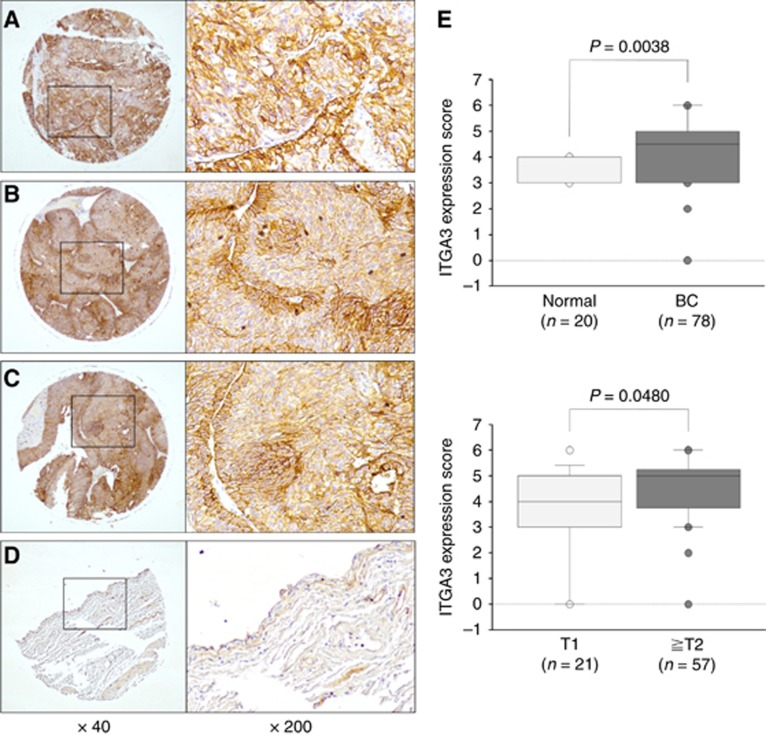
Figure 5.
IHC staining of ITGA3 in tissue specimens.(A) Strong positive staining in a tumour lesion (grade 2, T3N0M0), (B) strong positive staining in a tumour lesion (grade 1, T2N0M0), (C) strong positive staining in a tumour lesion (grade 2, T1N0M0), and (D) weak positive staining in normal bladder tissue. (E) ITGA3 expression scores in IHC staining; upper, ITGA3 expression in normal bladder tissues and BC; lower, correlation between ITGA3 expression and tumour status in BC.

