Figure 8.
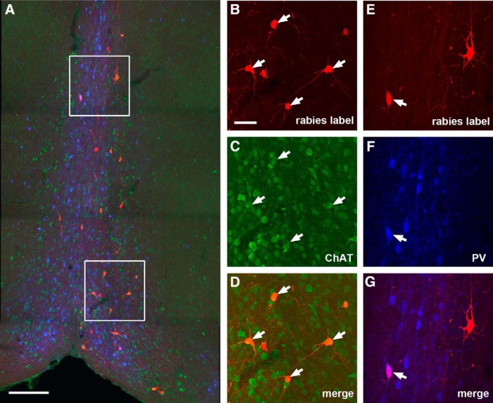
Cholinergic and GABAergic MS-DB inputs to dentate granule cells. A, An example MS-DB section image from a D1-Cre rabies tracing case. The red shows rabies-labeled neurons that are presynaptic to dentate granule cells, the green shows ChAT immunostaining, while the blue shows PV immunostaining. Scale bar, 200 μm. B–D, Enlarged view of the white box region at the top of A with arrows pointing to ChAT+ rabies-labeled septal neurons. E–G, Enlarged view of the white box region at the bottom of A with arrows pointing to a PV+ rabies-labeled septal neuron. The scale bar in B (50 μm) applies to B–G.

