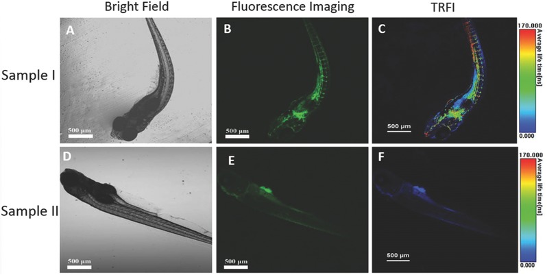
Figure 12.

Confocal fluorescence images of zebrafish: A–) zebrafish injected with CPy‐Odots; D–F) zebrafish noninjected with CPy‐Odots; (A, D) were bright field images; (B, E) were confocal fluorescence images recorded with 480–580 nm bandpass filters for CPy‐Odots upon excitation at 405 nm; (C, F) were lifetime images.
