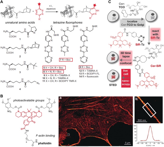
Figure 8.

Functional fluorophores using biomolecule‐based approaches. A) Protein labeling by inverse‐electron‐demand Diels–Alder cycloadditions. Structures of genetically encoded unnatural amino acids and tetrazine‐containing fluorophores. B) Photoactivatable phalloidin conjugate of 5‐carboxy‐NVOC2‐SiRhQ. a) Super‐resolution microscopy image of a COS‐7 cell stained with the phalloidin conjugate. b) Expanded image of the boxed region in (a), showing a protruding filopodial structure, and the line‐scan intensity across the filopodial structure in (b) (shown in black) and a Gaussian fit (red). C) Two‐step procedure for subcellular labeling of the Golgi apparatus in live cells; cells are treated first with Cer‐TCO, a trans‐cyclooctene‐containing ceramide lipid, and then reacted with the tetrazine fluorophore SiR‐Tz for 3D confocal and stimulated emission depletion (STED) super‐resolution microscopy. Reproduced with permission from Springer Nature68 (A) and Wiley‐VCH74, 77 (B, C).
