
Figure 7.
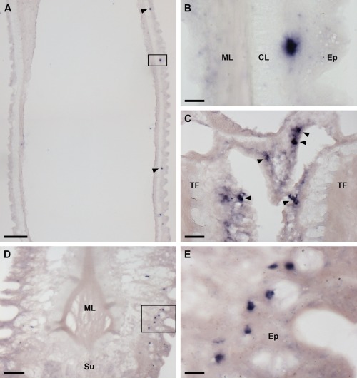
Localization of AruRGP precursor mRNA in tube feet of A. rubens using in situ hybridization. A: Longitudinal section of a tube foot showing three stained cells (arrowheads and rectangle) in the subepithelial layer of the podium. B: The region highlighted with a rectangle in A is shown here at higher magnification, with a stained cell located between the external epithelium and connective tissue layer. C: Stained cells (arrowheads) located in the subepithelial layer near to the base of adjacent tube feet. D: A group of stained cells (see rectangle) in the tube foot subepithelial layer just above the sucker. E: The region highlighted with a rectangle in D is shown here at higher magnification. CL, connective tissue layer; Ep, epithelium; ML, muscle layer; Su, sucker TF: tube foot. Scale bars = 100 μm in A; 10 μm in B; 25 μm in C; 50 μm in D; 10 μm in E.