
Figure 5.
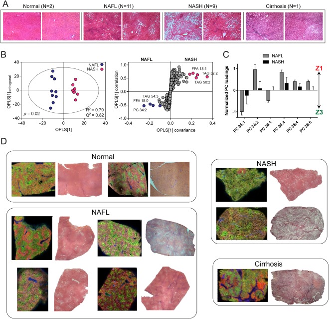
Lipid distributions across the human NAFLD spectrum. (A) H&E‐stained sections of human liver tissue for normal liver, NAFL, NASH, and cirrhosis. Original magnification: × 40. (B) Homogenized liver tissue was analyzed by LC‐MS, and the lipidomic profile was used to compare NAFL and NASH groups, revealing increased shorter chain TAGs and FFA(18:1) in NASH. (C) MALDI‐MSI was performed on frozen sections of tissue and quantified, revealing changes to zonation with NASH. (D) Example MS images of tissue covering the spectrum of NAFLD are shown alongside corresponding adjacent H&E sections. Spatial distributions for PC(32:0) (portal vein), PC(36:4) and PC(34:1) are denoted as blue, red, and green, respectively, for normal liver, NAFL, and NASH. Greater intensity of color reflects relative abundance. Whereas PC(36:4) and PC(34:1) are located in zones 1 and 3, respectively, in both normal and NAFL, there is partial to complete loss of zonation in NASH. Cirrhotic tissue, on the other hand, was marked by lipid changes to fibrotic and nodular regions (Supporting Information). Spatial distributions for lipids denoted in blue, red, and green for cirrhosis are PC(32:0), PC(34:1), and PC(34:2), respectively.