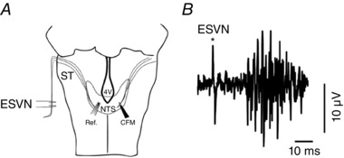
Figure 1. Schematic diagram of experimental set‐up.

A, carbon fibre microelectrodes (CFM) and reference electrodes (Ref.) were inserted in either side of the caudal nucleus of the solitary tract (cNTS). ST, Solitary Tract; 4V, 4th Ventricle. B, the location of the electrode in the cNTS was verified by recording evoked potential (multispike) responses from NTS neurons in response to electrical stimulation of the vagus nerve (ESVN).
