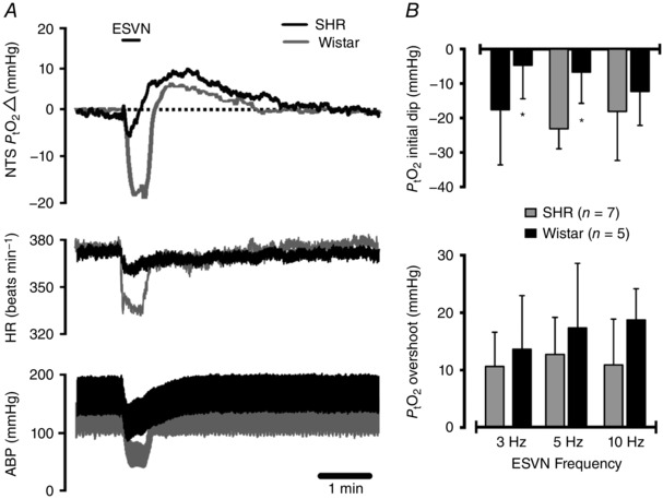
Figure 2. Measurements of tissue partial pressure of oxygen () in the caudal NTS of spontaneously hypertensive rats (SHRs) and Wistar rats with intact ganglionic transmission.

A, representative experimental trace showing the effect of electrical stimulation of the cut central end of the left vagus nerve (ESVN) on baseline arterial blood pressure (ABP), heart rate (HR) and . Functional activation of the NTS by ESVN caused a profound decrease in arterial blood pressure and heart rate as well as a biphasic change in characterized by an initial dip followed by an overshoot above baseline. B, group data showing the effect of ESVN at different stimulation frequencies. Data are presented as means ± SD. These changes were compared with control values using a two‐way ANOVA and the means compared with Fisher's LSD test.
