Figure 6.
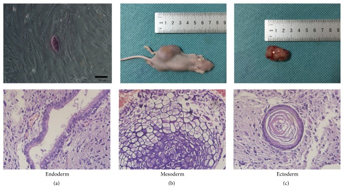
Identification of iPSCs. (a) The GFP+-iPSCs were purple which showed positive alkaline phosphatase staining. (b, c) Five weeks following injection, a 2.5 × 1.5 × 1.5 cm3 size tumor formed. Using hematoxylin and eosin staining, tumor tissue was noted to be derived from all three embryonic layers, including glandular epithelium (endoderm), cartilage (mesoderm), and cornified epithelium (ectoderm).

