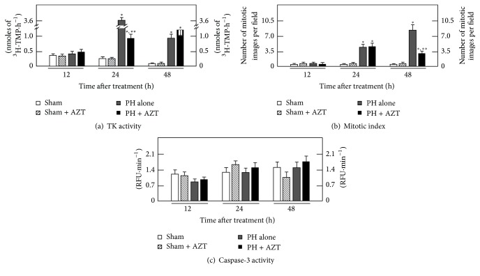
Figure 1.
Effects of AZT administration on some parameters indicative of cell proliferation and apoptosis at various times after 70%-PH. Results are expressed as mean ± SE for four independent determinations per experimental point for panel (a). The activity of TK expressed as nmoles of formed [3H]-TMP·h−1·mg−1 of cytosolic protein, in panel (b). The number of mitotic cells per microscopic field, as well as the cytosolic activity of caspase-3 (apoptosis) expressed as Relative Fluorescence Units (RFU)·min−1·mg−1 of protein (panel (c)). Symbols for the experimental groups at the bottom of each figure. Statistical significance: ∗p < 0.01 against Sham-operated (control) rats, and ∗∗p < 0.01 versus the PH group.

