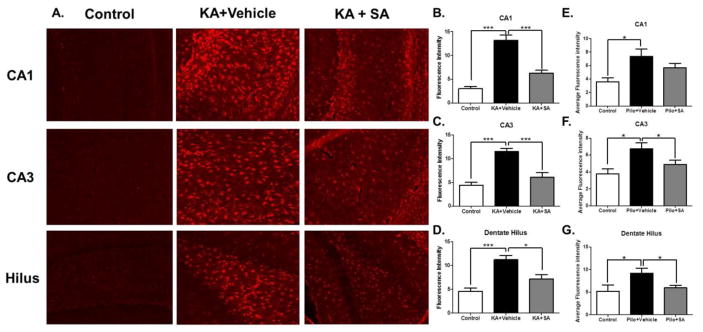
Figure 9. Pharmacological scavenging of gamma-ketoaldehydes attenuates astrogliosis in the KA model of TLE.
(A) Representative images of GFAP staining in brain regions taken 10 days after KA-induced SE. Quantification of the average fluorescence intensity in the KA model in (B) CA1 (C) CA3 and (D) hilus and in the pilo model (E) CA1 (F) CA3 and (G) hilus. *p< 0.05, ***p< 0.001 compared by one-way ANOVA, n =5–7/group.

