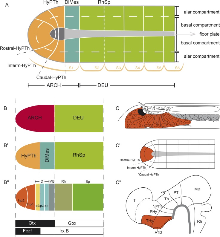
Fig 10. Genoarchitectonic model of the developing central nervous system (CNS) at the amphioxus 7-somite neurula stage.
(A) Summary of all identified anteroposterior (AP) and dorsoventral (DV) partitions of the neural plate of amphioxus. (B-B′′) Topological comparison of major molecular subdivisions between cephalochordates and vertebrates. (C,C′) Neural plate model highlighting the basal and alar plates of Rostral-hypothalamo–prethalamic primordium (Rostral-HyPTh) (orange) and the whole floor plate domain (gray) and its correspondence in a late larval stage (adapted from [90]). (C′′) Vertebrate neural tube highlighting the Terminal-Hypothalamic prosomere (orange) and the whole floor plate (grey).

