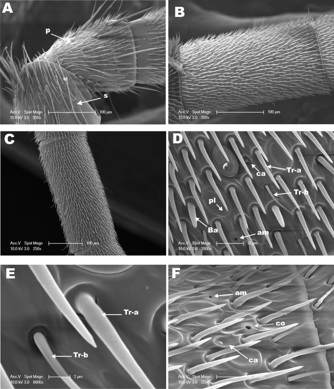
Fig 2. SEM from the dorsal side of the antenna of M. scutellaris queen.
(A) View of pedicel (P) and scape (S). Snapshots from (B) flagellomere 2 and (C) flagellomere 4. (D) Medial segment of flagellomere 5 highlighting showing sensilla trichodea a (Tr-a), trichodea b (Tr-b), sensilla basiconica (Ba), placodea (pl), campaniformia (ca) and ampullaceal (am). (E) Higher magnification of sensilla trichodea a (Tr-a) and trichodea b (Tr-b) from D. (F) Distal region of flagellomere 8 showing sensilla coeloconica (co), campaniformia (ca) and ampullaceal (am).

