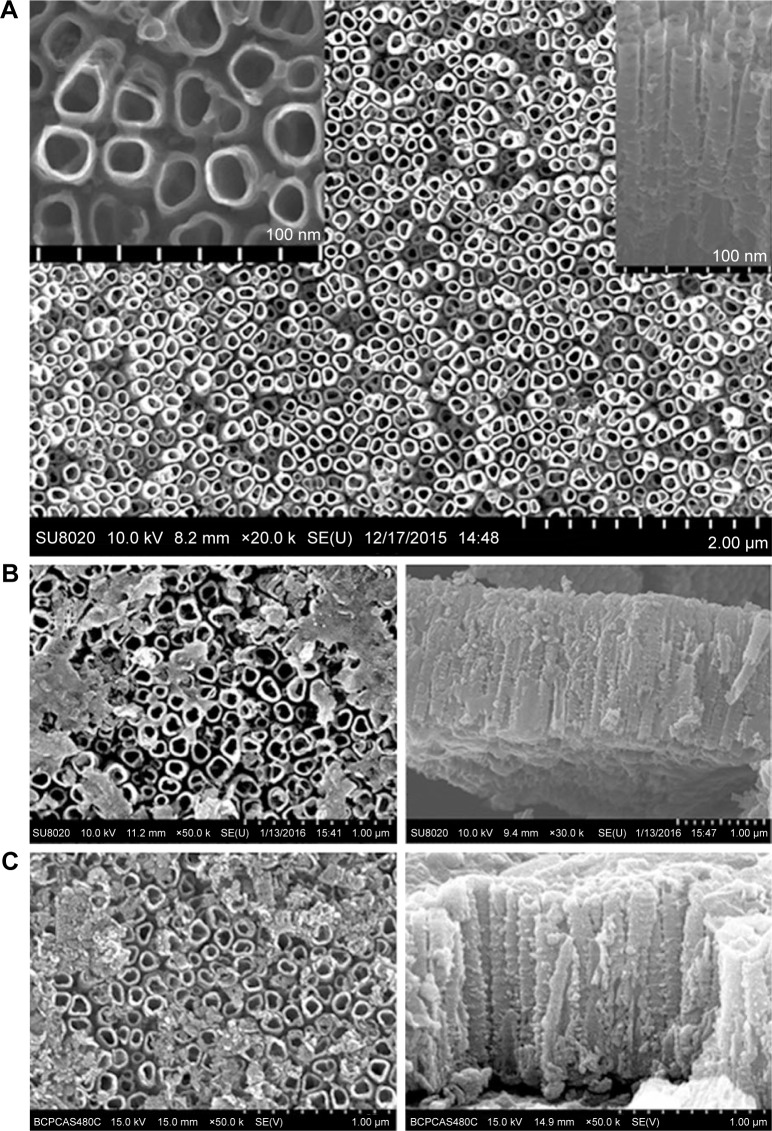
Figure 1.
FESEM micrographs of TNTs after annealing (A) including a high-magnification inset showing the pore diameter of 80 nm and cross-sectional view showing a length of approximately 2 µm; (B) MNA-TNTs showing the decoration of the TNTs with MNA flakes both in top view and in cross-sectional view; (C) GL13K-TNTs both in top view and in cross-sectional view.
Abbreviations: FESEM, field emission scanning electron microscope; MNA, metronidazole; MNA-TNTs, MNA-immobilized TNTs; TNTs, TiO2 nanotubes.

