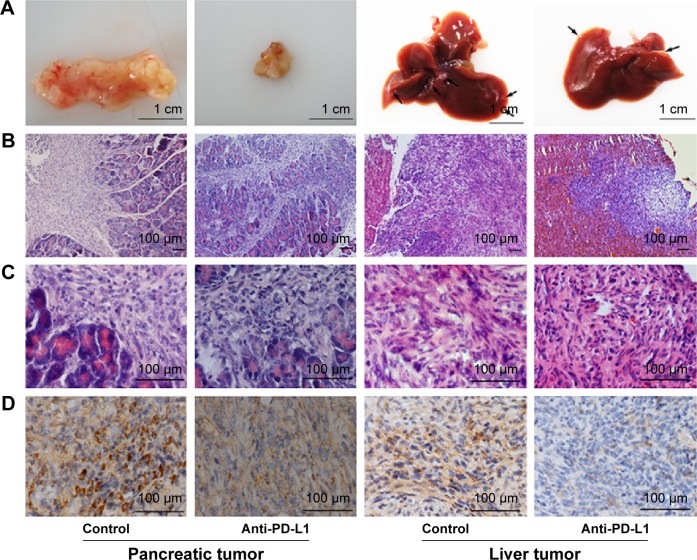
Figure 2.
Immunohistochemical analysis of PD-L1 expression in pancreatic cancer tissue and spontaneous liver metastases.
Notes: (A) Representative images of pancreatic tumor and spontaneous liver metastases. (B–C) Hematoxylin-eosin staining of pancreatic tumor tissue and spontaneous liver metastases at ×100 and ×400 magnification, respectively. (D) PD-L1 expression after injection with anti-PD-L1 antibody for 6 weeks in both pancreatic tumor tissue and spontaneous liver metastases at ×400 magnification.
Abbreviation: PD-L1, programmed death-ligand 1.

