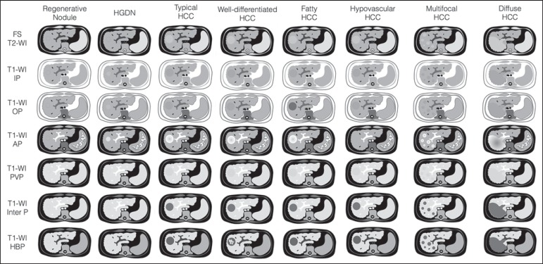
Figure 9.
Stereotypical simplified schematic representation, showing MRI features of cirrhotic nodules. In this schematic representation it is shown the appearance of common hepatocellular nodules in the cirrhotic liver, using a standard abdominal protocol. Abbreviations: HCC, hepatocellular carcinoma; HGDN, high grade dysplastic nodule; FS T2-WI, fat-suppressed T2-weighted image; T1-WI IP, T1-weighted in-phase image; T1-WI OP, T1-weighted out-of-phase image; T1-WI AP, post-contrast fat-suppressed T1-weighted image at the late arterial phase; T1-WI PVP, post-contrast fat-suppressed T1-weighted image at the portal-venous phase; T1-WI Inter P, post-contrast fat-suppressed T1-weighted image at the interstitial phase; T1-WI HBP, post-contrast fat-suppressed T1-weighted image at the hepatobiliary phase (with hepatobiliary contrast agent).

