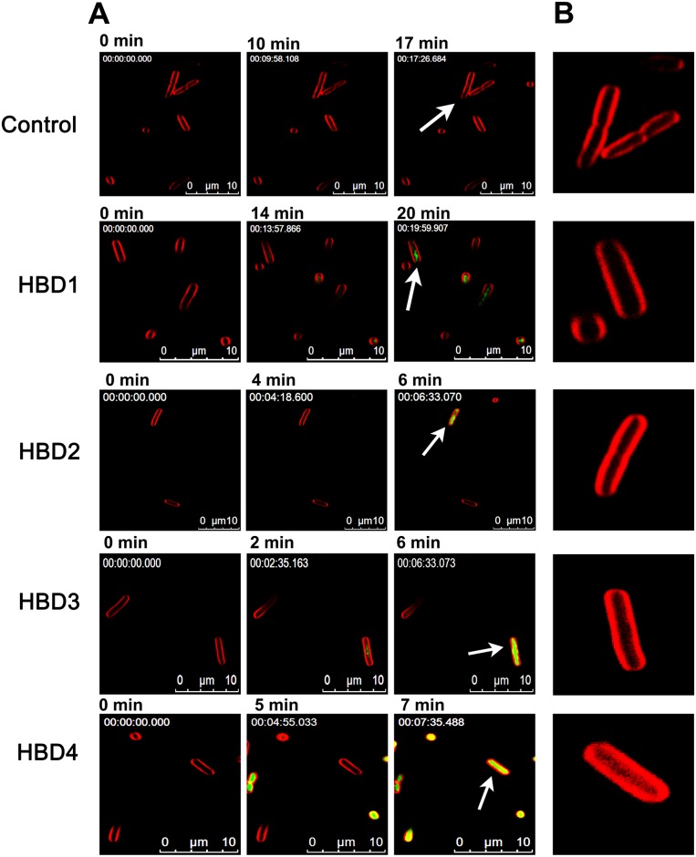
Fig 2. Effect of human β-defensins on the E. coli inner membrane.
(A), Intracellular accumulation of SYTOX green in defensin treated cells as function of time. The minute at which images were recorded is mentioned above the respective image. Numbers given in the upper left corner of the images represent the elapsed time (h:min:s:ms). (B), Morphological features of FM4-64 stained E. coli inner membrane of selected bacteria, as indicated by arrows.

