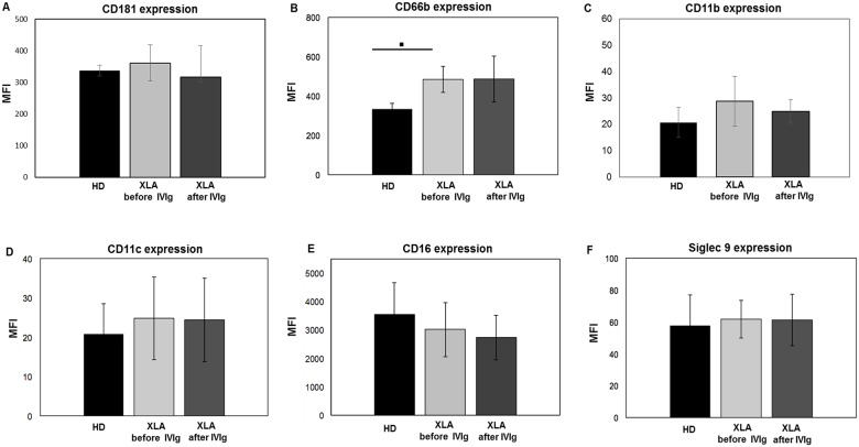
Fig 3. CD181, CD11b, CD11c and Siglec 9 expression on PMN from HD and XLA patients before and after IVIg infusion.
Whole blood samples were analyzed for the expression of CD181, CD66b, CD11b, CD11c, CD16 and Siglec 9 before and after IVIg infusion. The expression of all surface receptors was evaluated by performing a staining at 4°C for 30 min with specific fluorochrome-labeled antibody. Samples were washed, suspended in ice-cold PBS and analyzed by flow cytometry. XLA patients and HD show a similar CD181, CD11b, CD11c, CD16 and Siglec 9 expression, while CD66b expression was higher on PMN from XLA patients compared to HD (▪p = 0.001). After IVIg infusion, the expression of all receptors remained unaltered. Results are expressed as Mean Fluorescence Intensity. Histograms denote mean values and bars standard deviation. Statistical significance, determined by the nonparametric Mann Whitney test, is indicated as p value.

