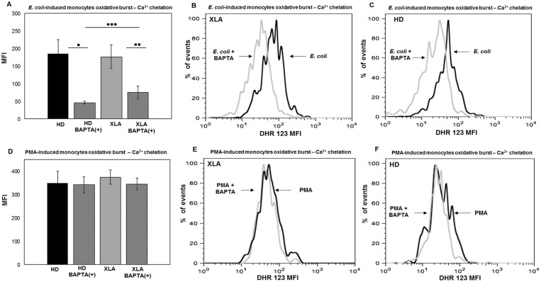
Fig 10. Monocytes oxidative burst—Ca2+ chelation.
Whole blood samples were pre-treated with 100 μM of the calcium chelator BAPTA-AM for 30 min and incubated in a water bath for 20 min at 37°C with opsonized E. coli (1-2x10⁹/ml) and PMA (1.62 mM). The conversion of DHR 123 to R 123 was used to evaluate the intracellular ROS production. FSC and SSC characteristics were used to identify the monocytes population. (A) BAPTA-AM treatment cause a stronger reduction of E. coli-induced oxidative burst in HD than XLA (▪▪▪p = 0.003). (A) The average reduction in HD was about 75% (▪p = 0.01) and it was about 50% in XLA patients (▪▪p = 0.02). (D) BAPTA-AM did not affect the oxidative burst induced by PMA both HD and XLA patients. Results are expressed as Mean Fluorescence Intensity. Histograms denote mean values and bars standard deviation. Statistical significance, indicated as p value, was determined by the nonparametric Mann-Whitney test and Wilcoxon Signed Rank test. (B and C) It shown monocytes E. coli-induced oxidative burst in a representative HD and XLA patient, black peak BAPTA-AM(-) and gray peak BAPTA-AM(+). (E and F) It shown monocytes PMA-induced oxidative burst in a representative HD and XLA patient, black peak BAPTA-AM(-) and gray peak BAPTA-AM(+).

