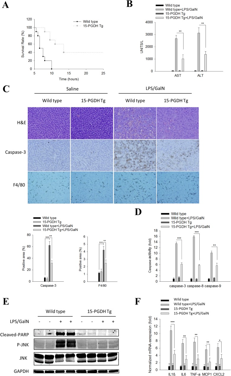
Fig 1. Liver specific 15-PGDH expression protects mice against LPS/GalN-induced acute liver inflammation and tissue damage.
Wild type and 15-PGDH Tg mice were intraperitoneally injected with LPS (60ng/g body weight) plus GalN (800μg/g body weight). The mice were followed for survival or sacrificed at 5h after LPS/GalN injection to collect serum and liver tissue samples.(A) Survival curve of mice after LPS/GalN administration (n = 10). (B) Serum ALT and AST levels (n = 5). (C) Representative images of liver tissue sections (200×): H&E stain (upper panel), Caspase-3 IHC (mid panel), F4/80 IHC (lower panel). Quantified IHC results was shown below (n = 5). (D) Caspase-3, 8, and 9 activities in liver tissue homogenates (n = 3). The data were shown as fold changes compared to wild type mice. (E) The protein levels of apoptotic signaling molecules (Cleaved PARP, JNK, P-JNK) in liver tissue homogenates. GAPDH was used as loading control. (F) mRNA levels of pro-inflammatory cytokines (IL1β, IL6, TNF-α, MCP1 and CXCL2) in liver tissue homogenates (n = 3); the results are shown as fold changes compared to wild type mice. The quantitative data presented in this figure are expressed as means ± SE (*p<0.05, **p<0.01, ***p<0.001).

