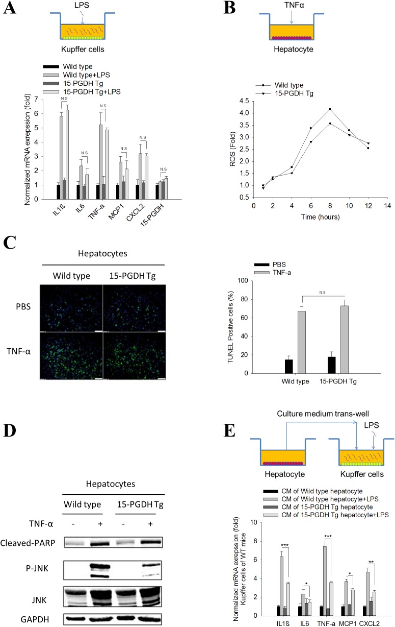
Fig 2. 15-PGDH expression in hepatocytes regulates Kupffer cell cytokine production.
Kupffer cells and hepatocytes were isolated and cultured separately. LPS (10ng/ml) was used to elicit Kupffer cell inflammatory response. TNF-α (25ng/ml) plus ActD (0.4μg/ml) was used to induce hepatocyte apoptosis. (A) mRNA level of pro-inflammatory cytokines (IL1β, IL6, TNF-α, MCP1 and CXCL2) and 15-PGDH in Kupffer cells after LPS treatment. The results are shown as fold changes compared to Kupffer cells isolated from wild type mice. (B) Accumulation of reactive oxygen species (ROS) in hepatocytes after TNF-α/ActD treatment. ROS was measured by dichlorofluorescin fluorescence assay and shown as fold change compared to untreated hepatocytes. (C) Hepatocyte apoptosis induced by TNF-α/ActD treatment. Apoptotic hepatocytes were stained by TUNEL assay. Representative images are showed in the left panel. Quantified results are showed in the right panel. (D) The protein levels of apoptotic signaling molecules (cleaved PARP, JNK, P-JNK) in hepatocytes after TNF-α/ActD treatment. GAPDH was used as loading control. (E) mRNA level of pro-inflammatory cytokines (IL1β, IL6, TNF-α, MCP1 and CXCL2) in WT Kupffer cells treated with hepatocyte CM followed by LPS stimulation. The results are presented as fold changes compared to Kupffer cells treated with CM of wild type hepatocytes. The quantitative data presented in this figure were obtained from three independent experiments and are expressed as means ± SE (*p<0.05, **p<0.01, ***p<0.001; N.S denotes no statistical significance).

