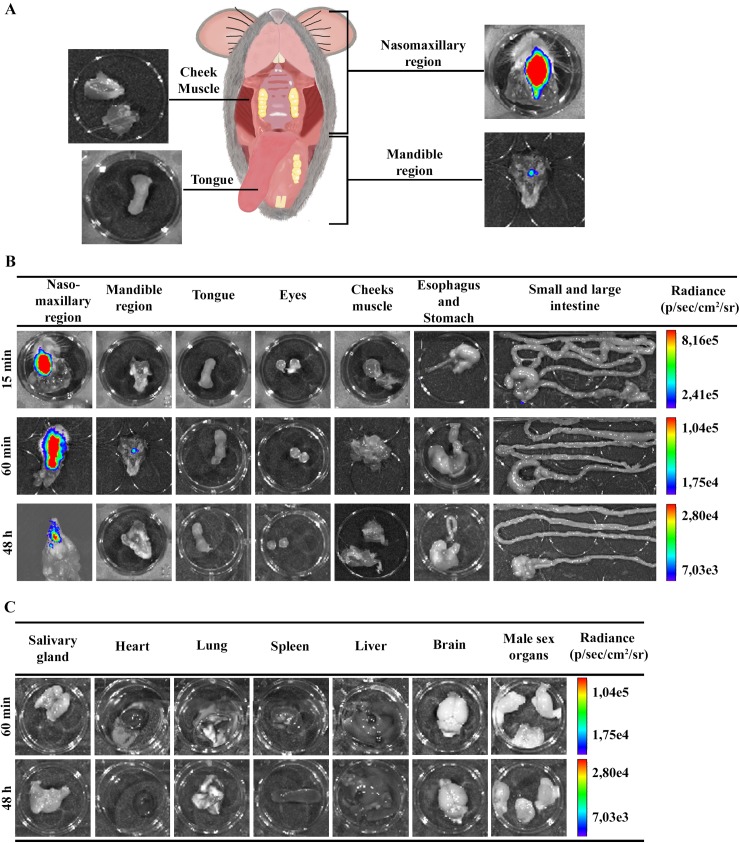
Fig 3. Ex vivo evaluation of dissected organs and tissues by bioluminescent imaging.
Male BALB/c mice were infected in the oral cavity (OI) with 1x10⁶ trypomastigotes forms of T. cruzi expressing luciferase (Dm28c-luc). After 10 min of D-luciferin i.p administration (150 mg/kg), organs were harvested and images were captured using an IVIS Lumina II system. (A) Schematic picture for anatomic localization of organs and tissues analyzed. Nasomaxillary region includes all tissues from regions of the nose, nasal cavity and upper region of the oral cavity with exception of the cheek muscle. Mandible region includes all tissues of the mandible and the lower region of the oral cavity, with exception of the tongue (B and C). Ex vivo bioluminescence imaging from selected organs and tissues at 15 min (n = 3), 60 min (n = 4 in the nasomaxilary region; n = 2 in other organs) and 48 hours (n = 5) post-infection. The scale bar for radiance (right) was correlated with the signal intensity, where red indicates higher signal and blue indicates a lower signal. Maximum and minimum signals are indicated at the top and bottom of the scale bar, respectively.

