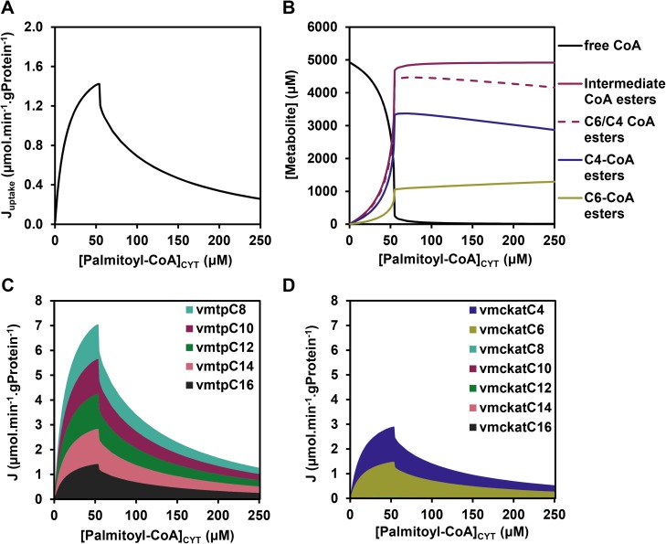
Fig 2. Simulated steady-state fluxes and concentrations in the mFAO model.
(A) The effect of cytosolic palmitoyl-CoA concentration ([palmitoyl-CoA]CYT) on the steady-state flux. The steady-state uptake flux of palmitoyl-CoA (Juptake) is plotted (calculated as the steady-state flux of palmitoyl-carnitine through CACT, i.e. the uptake of palmitoyl-carnitine into the mitochondria), in contrast to van Eunen et al. 2013 [8] in which the NADH flux was plotted. The two are uniquely linked with the latter being 7-fold higher. (B) The steady-state concentrations of free CoA (identical to data from [8]), the sum of all intermediate CoA esters, the sum of all C4- and C6 CoA esters and the subset of C4- and C6 CoA esters (new results). (C-D) Distribution of steady-state fluxes (J) of different chain-length substrates through MTP and MCKAT, respectively. Only the chain lengths which are converted by these enzymes (cf. Fig 1) are included.

