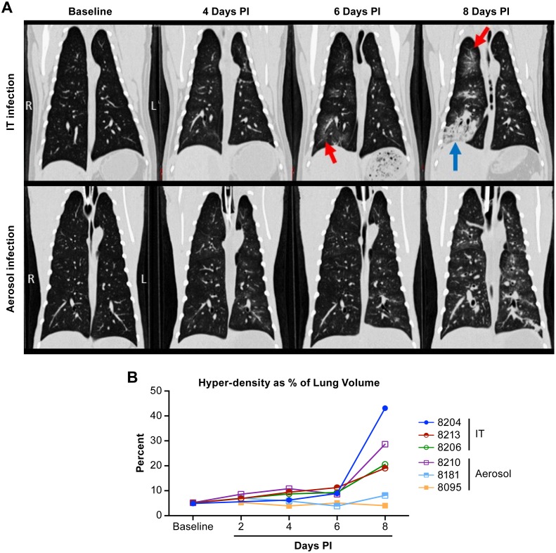
Fig 4. CT analysis of the lungs of NiV infected AGMs.
Examples of temporal analysis of changes within the lungs of AGMs infected by intratracheal or aerosol inoculation (A). In animals infected by the IT route, images shows a vascular pattern of infiltration extending to adjacent alveoli, manifested as radiating ground glass opacities (indicated by red arrows), progressing by day 8 to diffuse lobar consolidation (blue arrows) that tends to extend from cranial to caudal in severity. In animals infected by aerosolization of small particles, diffuse progressive thickening of airway walls and adjacent interstitium are typically observable within 6 days of infection, which ultimately extends into the alveoli, uniformly throughout the lungs. Where IT inoculation tends to induce some amount of consolidation, typically in caudal lobes, infection by small particle aerosol appears to cause disseminated pulmonary changes throughout all lobes. Quantification (B) of lung volume demonstrates that animals lost up to 45% of their total lung volume with all three of the animals infected by IT inoculation losing 18% or greater of their lung volume.

