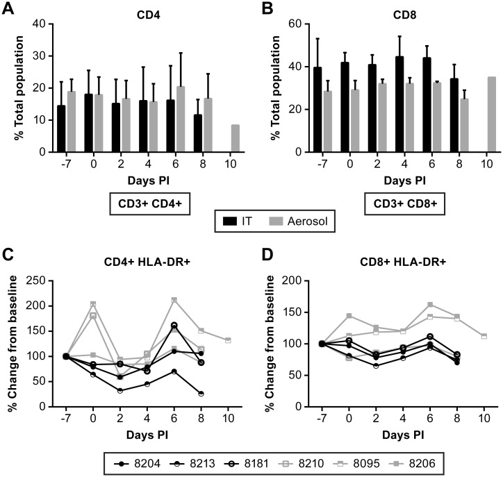
Fig 8. Evaluation of T cell responses in NiV infected AGM over the course of disease.
Flow cytometric analysis of changes in CD4+ (A) and CD8+ (B) populations. Evaluation of changes in activated T cell populations was performed using HLA-DR as a marker for activation and was referenced to a pre-infection bleed. No significant changes were noted in activated CD4+ T cells (C), but two animals had nominal increased in activated CD8+ T cells (D) relative to other animals. Animals were infected with NiV by IT (black) or aerosol (gray) inoculation.

