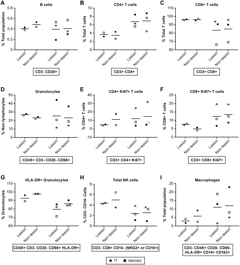
Fig 9. Evaluation of immune cell population in the lung collected at necropsy.
Comparisons of immune cell populations in “lesion” and “non-lesion” samples collected at necropsy from animals infected by IT (●) or aerosol (■) inoculation. Markers are consistent for individual animals. IT inoculated: 8204 (●), 8213 (half-filled ○) and 8181 (○); Aerosol inoculated: 8210 (□), 8095 (half-filled □) and 8206 (■). Represented are data for (A) B cells, (B) CD4+ T cells, (C) CD8+ T cells, (D) Granulocytes, (E) Ki67+ CD4+ T cells, (F) Ki67+ CD8+ T cells, (G) HLA-DR+ Granulocytes, (H) Total NK cells and (I) Macrophages. These data suggest that CD4+ cells are elevated in aerosol inoculated animals relative to IT inoculated while activated granulocytes (HLA-DR+) and NK cells appear somewhat elevated in IT inoculated animals. Statistical analyses were not performed due to limited group size.

