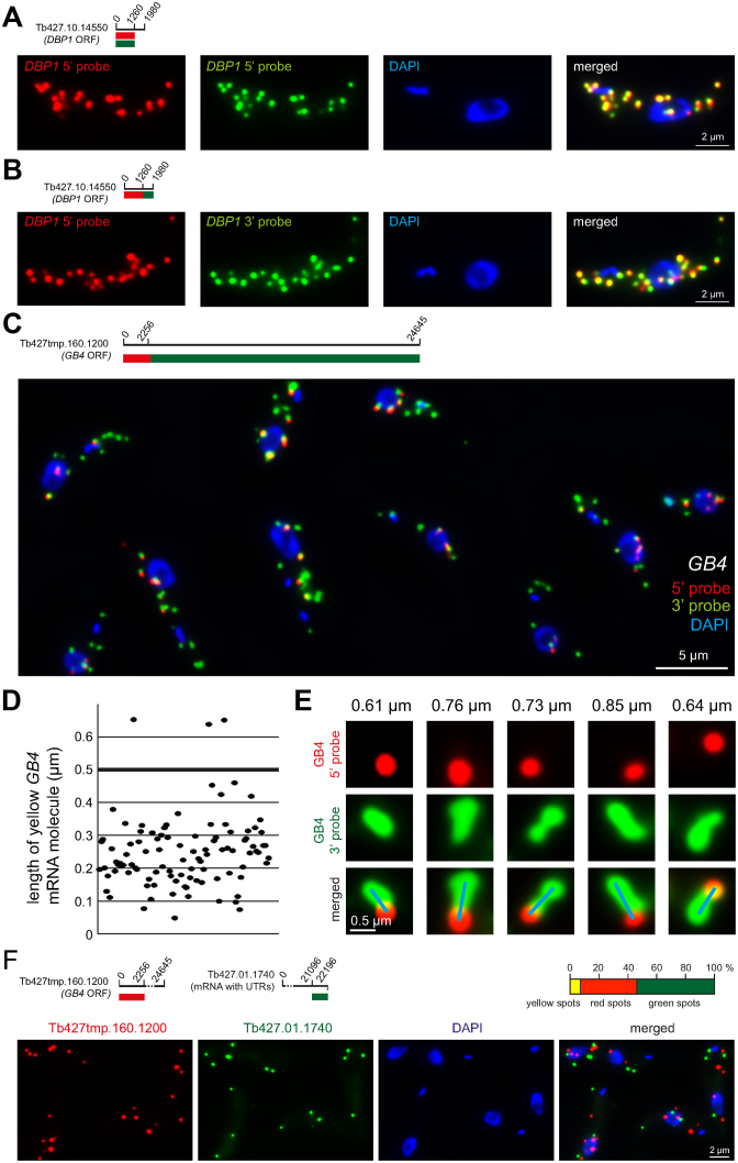
Figure 2.
Technical controls and the definition of a yellow spot. (A) Red and green fluorescence in situ hybridization (FISH) probes were designed to anneal to the same mRNA sequence of an average-sized mRNA (Tb427.10.14550). The signals from the two probes showed a strong overlap. One representative cell is shown; more cells are shown in Supplementary Figure S2. (B) Red and green FISH probes were designed to anneal to adjacent mRNA sequences of the same, average-sized mRNA. The signals from the two probes again showed a strong overlap. One representative cell is shown; more cells are shown in Supplementary Figure S3. (C–E) Trypanosome cells were simultaneously probed with the 5΄ probe (red) and the 3΄ probe (green) specific for the GB4 mRNA (Tb427tmp.160.1200) (details on top left of C). Together, these probes cover the entire 24 645 nucleotides of the GB4 open reading frame: the 5΄ probe binds to the 2256 nucleotides at the 5΄ end and the 3΄ probe to the remaining 22 389 nucleotides. (C) One representative Z-stack projection image with merged fluorescence channels is shown. (D) The length of 100 yellow GB4 mRNA molecules was measured from Z-stack projection images. In total, 97% are less than 0.5 μm in length (D). (E) Example images of very extended GB4 mRNA molecules; the length is indicated on top and the blue line shows how the length measurement was performed (from the middle of the ‘red circle’ to the middle of the most distant ‘green circle’ at the end of the green string). These very long mRNA molecules are rare and never longer than 1.0 μm. (F) Trans-probing: dual colour mRNA FISH using probes antisense to two different mRNAs (details on top left). The percentage of red, green and yellow spots was quantified from 152 cells (top right). The average cell had 7.3 ± 2.8 mRNA molecules. A representative image is shown (bottom).

