Figure 1.
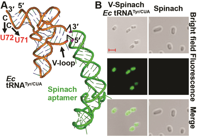
Expression of a V-Spinach tRNA in E. coli. (A) The Spinach aptamer (in green) is fused to the V-loop of E. coli (Ec) tRNATyr/CUA (in orange), harboring the amber-reading anticodon 5΄-CUA-3΄. The drawing of the Spinach aptamer is based on the crystal structure of the Fab BL3-6-bound aptamer (29). The sequence of the Spinach aptamer is as described (15) and it is covalently joined to the V-loop between C47:2 and A47:3 as shown by two black up arrow icons. The U71 and U72 substitutions in Ec tRNATyr are shown by downward arrows, which replace G1-C72 and G2-C71 base pairs with G1-U72 and G2-U71, respectively. (B) Fluorescence microscope images of E. coli cells expressing the amber suppressor form of V-Spinach tRNATyr versus cells expressing the Spinach stand-alone. DIC images and scale bars (each representing 2 μm) are shown. On average, when examined in liquid cultured media, 96.4 ± 0.1% of cells expressing the V-Spinach tRNA showed the GFP-like fluorescence (n = 100).

