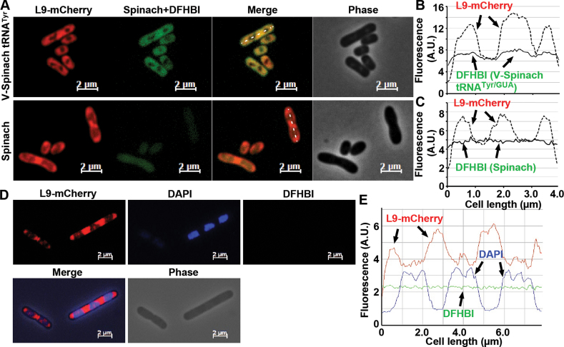
Figure 4.
Localization of V-Spinach tRNATyr in living E. coli cells relative to L9-mCherry ribosomes. (A) E. coli JM109 cells, expressing L9-mCherry from the chromosome and V-Spinach tRNATyr/GUA (top) or the stand-alone Spinach (bottom) from pKK223-3 (induced with IPTG), were observed under microscope as described in Materials and Methods. (B and C) Lengthwise intensity scans of mCherry (dotted line) and Spinach fluorescence (solid line) of a representative cell from the two strains. (D) E. coli JM109 cells, harboring no plasmid, were imaged for L9-mCherry (left) and with DFHBI (right) and stained with DAPI (middle). (E) Lengthwise intensity scans of mCherry (red line), Spinach fluorescence (green line), and DAPI stain (blue line) of a representative cell.

