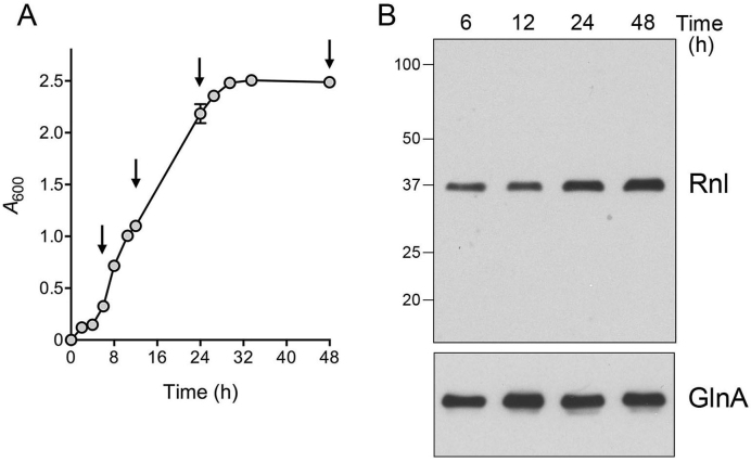
Figure 1.
DraRnl is constitutively expressed in D. radiodurans. (A) Cells were inoculated from an overnight culture into 1× TGY medium to attain A600 of 0.05 and the culture was incubated with constant shaking at 30°C. Growth was monitored by plotting A600 as a function of time post-inoculation. Aliquots were removed at the times specified by arrows (6, 12, 24 and 48 h). (B) Western blotting. Aliquots of cells were normalized to A600 of 1.0, pelleted and resuspended in 10 mM sodium phosphate (pH 7.5). Cells were then lysed in SDS loading buffer, resolved by 10% SDS-PAGE, and transferred to a nitrocellulose membrane. An immunoblot with anti-DraRnl antibody is shown in the top panel. The positions and sizes (kDa) of marker polypeptides are indicated on the left. An immunoblot with anti-GlnA antibody is shown in the bottom panel.

