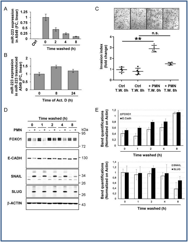
Figure 4.
Engulfed miR-223-3p is quickly decayed in recipient cells after donor cell removal. (A) Time-dependent decay of ex-miRNA-223-3p expression in A549 cells. Following overnight co-culture with PMN, A549 cells were harvested at the indicated time periods after PMN removal. The level of miR-223-3p was measured by RT-qPCR and normalized using U6 snRNA. Results are representative of three biological replicates, ‘centre values’ as mean and error bars as s.d. (B) RT-qPCR analysis of miRNA-223-3p expression in stably miR-223 transduced A549 cells treated with actinomycin D (Act. D) at 10 μg/ml for the indicated period of time. Spikes-in were used for normalization (see the ‘Materials and Methods’ section). (C) In vitro invasion assay of A549 cells post washes. A549 cells co-cultured with (+PMN) or without PMN (Ctrl) overnight were collected at the indicated time post initial washes (Time Washed, T.W.) and seeded, in the upper part of transwells. The number of cells attached to the bottom of a Matrigel-coated membrane after 16 h was quantified after crystal violet staining. Data represent the quantification of five biological replicates, ‘centre values’ as mean and error bars as s.d. (D) Immunoblot analysis of FOXO1 and EMT marker expression levels upon PMN removal from A459 cells co-cultured with (+) or without (−) PMN. β-ACTIN served as reference for loading. (E) Quantifications of gels from three independents experiments are presented. * for P < 0.05, ** for P < 0.01.

