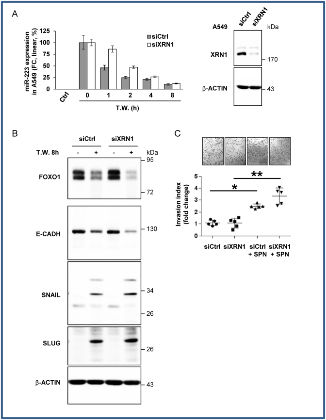
Figure 5.
XRN1 regulates ex-miRNA decay in recipient cells (A) Relative quantification analysis of ex-miRNA-223-3p in A549 cells. siXRN1-transfected A549 cells co-cultured with PMN overnight were harvested at the indicated periods of time post PMN removal (Time Washed, T.W.). Results are representative of three biological replicates. In the right panel, immunoblot analysis of XRN1 expression. b-ACTIN served as an equal loading control. (B) Immunoblot analysis of FOXO1 and EMT marker expression levels. β-ACTIN served as an equal loading control. (C) In vitro invasion assay of siXRN1-transfected A549 cells. A549 cells co-cultured with SPN of PMN, produced in serum-free medium, were seeded in the upper part of transwells. The number of cells attached to the bottom of a Matrigel-coated membrane after 16 h was quantified after crystal violet staining. Data represent the quantification of five biological replicates, ‘centre values’ as mean and error bars as s.d. * for P < 0.05, ** for P < 0.01.

