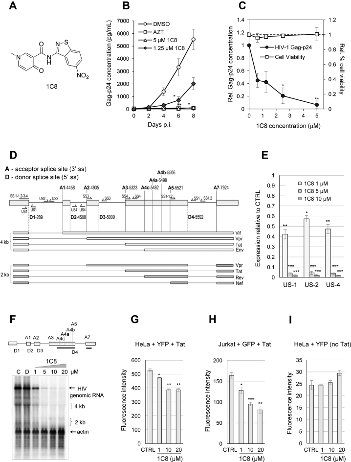
Figure 1.
Effect of 1C8 on HIV-1 expression. (A) Chemical structure of 1C8. (B) Effect of 1C8 on HIV-1 replication in PBMCs. Assay monitoring HIV-1 BaL virus replication over a period of eight days post-infection (p.i.) as measured by Gag-p24 antigen by ELISA (n > 3, 3–4 donors). PBMCs were infected with HIV-1 BaL (MOI < 0.01) and treated on days 0 and 4 post-infection with DMSO, AZT (3.74 μM), or 1C8 at the concentrations indicated. Error bars indicate standard error of the mean (SEM) of replicate wells from an independent experiment. (C) Effect of increasing concentrations of 1C8 on cell viability in PBMCs. Culture supernatants were measured for Gag-p24 antigen by ELISA and expressed relative to DMSO treatment (n ≥ 3, *P ≤ 0.05, **P ≤ 0.01 and ***P ≤ 0.001). The effect of the compounds on cell viability was measured by trypan blue exclusion as a percentage of total cells and expressed relative to percent cell viability with DMSO treatment. (D) Map of donor and acceptor splice sites (5΄ss and 3΄ss, respectively) used for the production of the major HIV mRNAs. The main viral proteins produced from each mRNA are indicated. (E) Quantitative RT-PCR assays from HeLa-HIV cells treated with 1C8. Histograms depicting the impact of 1C8 on amplicons derived from unspliced segments of HIV-1 transcripts. The intensity of products was normalized relative to the amount produced in the controls (which corresponds to an arbitrary unit value of 1). (F) Northern analysis of HIV-1 transcripts following treatment with various concentrations of 1C8 for 24 h. The HIV probe is indicated by a black line below the HIV map. A β-actin probe was used to reveal loading variations. Positions of 4 and 2 kb species are indicated. (G) HeLa cells were co-transfected with a HIV-YFP plasmid and a CMV-Tat plasmid in the presence of the indicated concentrations of 1C8. Total YFP fluorescence of YFP positive cells was measured 36 h post-transfection. (H) Jurkat cells were co-transfected with a HIV-YFP plasmid and a CMV-Tat plasmid in the presence of the indicated concentrations of 1C8. Total YFP fluorescence of YFP positive cells was measured 36 h post-transfection. (I) HeLa cells were transfected with the HIV-YFP plasmid in the presence of the indicated concentrations of 1C8. Total YFP fluorescence of YFP positive cells was measured 36 h post-transfection. In all cases, asterisks represent significant P values (two-tailed Student's t test) comparing the means between 1C8-treated samples and their respective controls; *P < 0.05, **P < 0.01 and ***P < 0.001.

