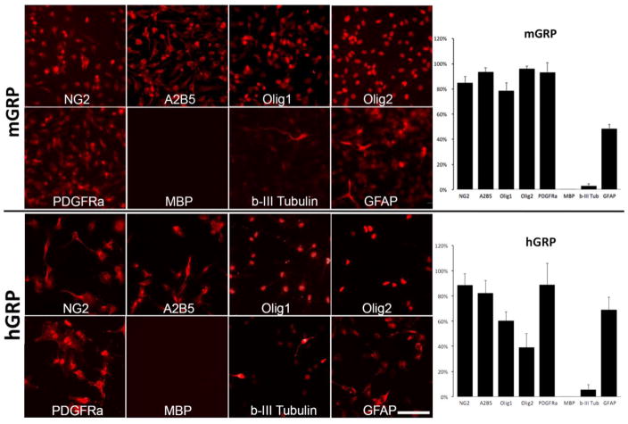
Figure 1. Phenotypic characterization of mouse and human GRP cells prior to transplantation.
Mouse and human GRPs were derived from mid-gestation (day 12; week 22 respectively) whole forebrain and immunostained within three days of culture. for NG2, A2B5, Olig1, Olig2, PDGFRa, MBP, b-III Tubulin, GFAP. Scale bar=100 μm applies to all images. Graphs show quantification of cells positive for specific antigens.

