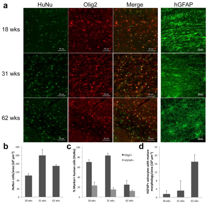
Figure 8. Temporal characterization of cell fate in hGRP-grafted shiverer mice.
(a) Engrafted human cells 18, 31, and 62 weeks after transplant into neonatal shiverer mice were stained for HuNA and counterstained with Olig2 or hGFAP (50 μm and 20 μm scale bar, respectively). (b) Quantification of the number of human cells, identified by HuNa, at successive time points after transplantation in the corpus callosum. (c) Quantification of human cell fate 18, 31, and 62 weeks after transplantation in the corpus callosum (Olig2 and hGFAP). (d) Quantification of hGRP-derived mature astrocytes in the corpus callosum.

