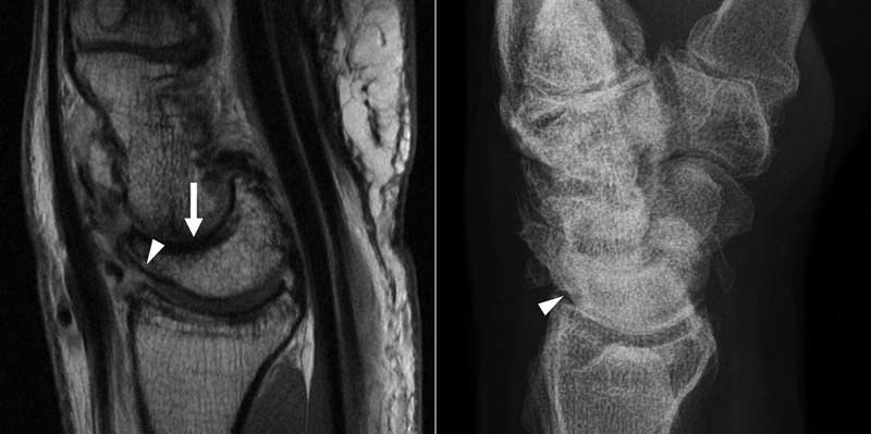
Fig. 1.

A 71-year-old man with SLAC arthritis. (A) Sagittal fast spin-echo magnetic resonance imaging demonstrates high grade to full thickness chondral loss at the dorsal margin of the radiolunate joint (arrowhead). Full thickness chondral loss is also seen at the capitolunate joint (arrow). (B) Corresponding radiograph demonstrates a normal radiolunate joint.
