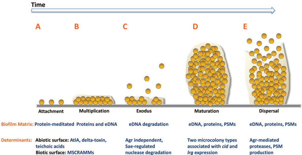
Figure 1. Model of Staphylococcus aureus biofilm development.
S. aureus biofilm development is described in five stages: A) attachment, B) multiplication, C) exodus, D) maturation, and E) dispersal. A. S. aureus cells attach to abiotic or biotic surfaces via hydrophobic interactions or MSCRAMMs, respectively. B. After cells attach, the biofilm develops into a confluent ‘mat’ of cells composed of an eDNA and proteinaceous matrix. C. Upon reaching confluency, a period of mass exodus of cells occurs in which a subpopulation of cells is released from the biofilm via Sae-regulated nuclease-mediated eDNA degradation to allow for the formation of three-dimensional microcolonies. D. Microcolonies form from distinct foci of cells that have remained attached during the exodus stage. This stage is characterized by rapid cell division that forms robust aggregations composed of proteins including PSMs and eDNA. E. Activated Agr-mediated quorum sensing initiates biofilm matrix modulation and dispersal of cells via protease activation and/or PSM production. AtlA, autolysin A; MSCRAMM, microbial surface components recognizing adhesive matrix molecules; eDNA, extracellular DNA; PSM, phenol soluble modulins; Agr, accessory gene regulator.

