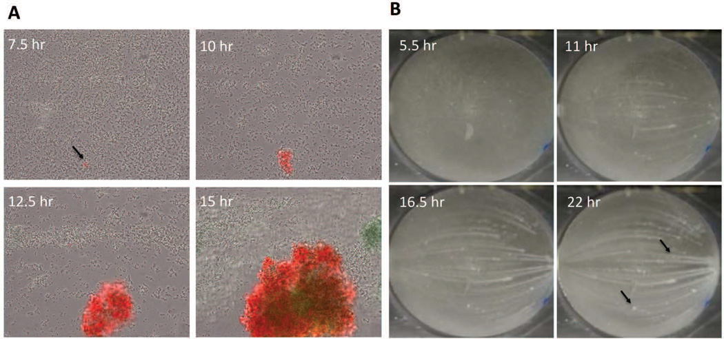
Figure 3. Microcolony initiation.
A) S. aureus cells containing an lrgAB::gfp promoter fusion plasmid were inoculated into a Bioflux1000 microfluidics system and allowed to form a biofilm over a time-course experiment in which epifluorescence images were acquired at regular time points. Shown are images collected at regular intervals after the initiation of medium flow. Note the emergence of the microcolony originating from what appears to be a single (or relatively few) lrgAB-expressing (green) cells. (B) Macroscopic images of S. aureus biofilm grown in an FC flow-cell system. Shown are images collected at 5.5, 11, 16.5, and 22 hrs after the initiation of medium flow. Note the emergence of microcolonies from a basal layer of cell starting at 5.5 hrs, as well as the presence of streaking downstream of most (but not all; see arrows) of the microcolonies.

