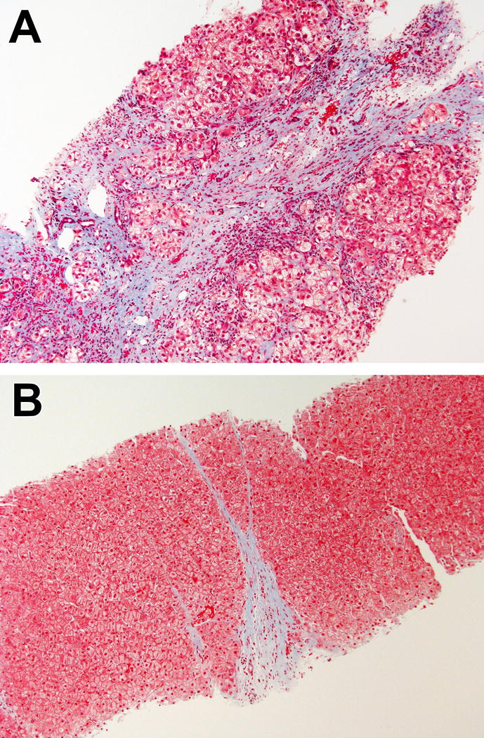
Figure.

Examples of progressive (A) and regressive (B) fibrosis phenotypes. Both cases were staged as cirrhotic (Ishak fibrosis stage 6). In (A), the fibrous band is wide, with uneven staining and shows disruption of the adjacent parenchyma. In (B), the fibrous band is thin, with homogeneous staining and sharp borders. (Masson trichrome, 100× for both).
