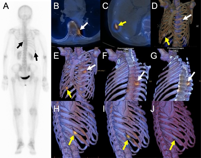
Fig. 5.
Whole-body scintigraphy (a), 2D SPECT/CT fusion (b and c), 3D SPECT/CT fusion using volume-rendered SPECT images (d) or image data projection of bone SPECT onto 3D volume-rendered CT images without (e, h, and j) or with clip-plane editing (f, g, and i) in a breast cancer patient with co-existence of bone metastasis and fracture in the ribs. Osteolytic change is clearly seen in 2D SPECT/CT (b) and our 3D SPECT/CT (f and g) images in the proximal site of the left eighth rib (white arrows). By contrast, a fracture line is shown at the hypermetabolic site in 3D SPECT/CT fusion (h–j)

