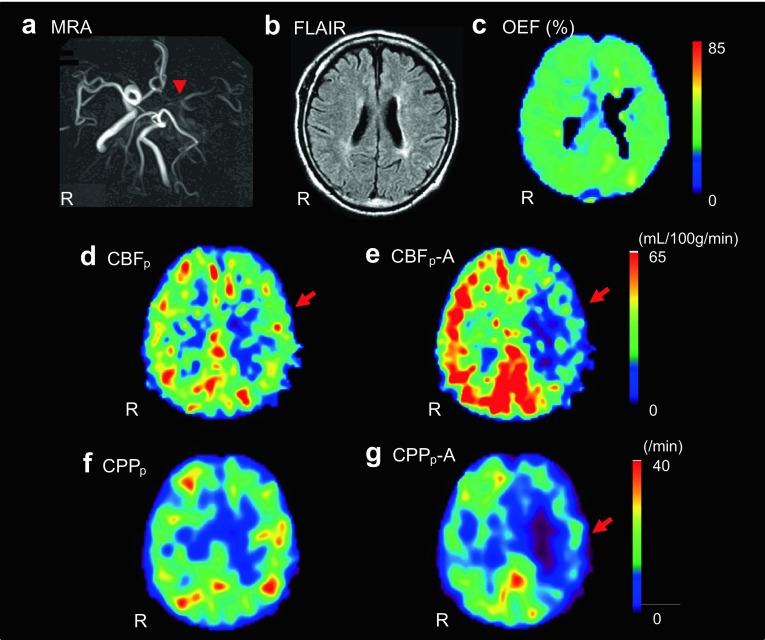
Fig. 6.
A representative case from this study. The patient had left ICA occlusion on MRA (a). Arrowhead shows the occlusion of ICA. MRI FLAIR image showed no significant ischemic region in the MCA territory (b). Although OEF image showed no significant increase in the left cerebral hemisphere (c), baseline CBFp showed a tendency of decrease in the MCA territory (d). ACZ challenge test showed significant decrease in CBFp (e) and CPPp (f baseline, g after ACZ) in the affected region. The study indicated significant hemodynamic impairment because of the significant decrease in CVR and CPP, and the patient underwent EC–IC bypass surgery. CBFp-A and CPPp-A means pixel-by-pixel calculated CBF and CPP after ACZ. R on each panel represents right side of the patient. Red arrows show affected side of MCA territory

