Figure 3.
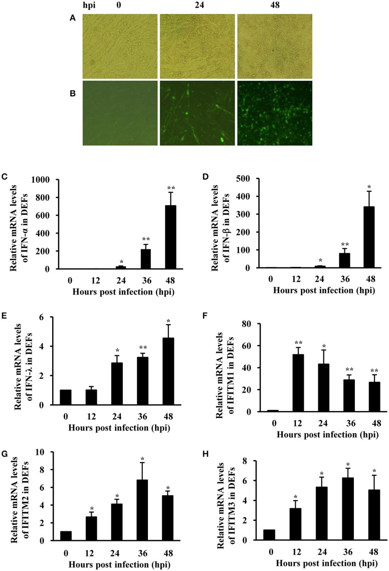
IFNs and IFITMs are significantly induced in DEFs after ATMUV infection. DEFs were infected with or without ATMUV at a MOI of 1.0 and harvested at 0, 12, 24, 36, and 48 hpi, respectively. (A) CPE feature was recorded at 0, 24, and 48 hpi. (B) Viral antigens were detected by IFA in DEF cells at 0, 24, and 48 hpi. qRT-PCR analysis was performed to examine the mRNA expression of duck type I and type III IFNs (C–E) and IFITM1, 2, 3 (F–H). The mRNA levels were normalized to the endogenous β-actin level and the expression at 0 hpi was set to 1.0. Expression at 12–48 hpi was compared to its expression at 0 hpi. Plotted are the average levels from three independent experiments with three replicates per experiment (means ± SD). Statistical significance was determined by one tail Student's t-test analysis. *P < 0.05, **P < 0.01.

