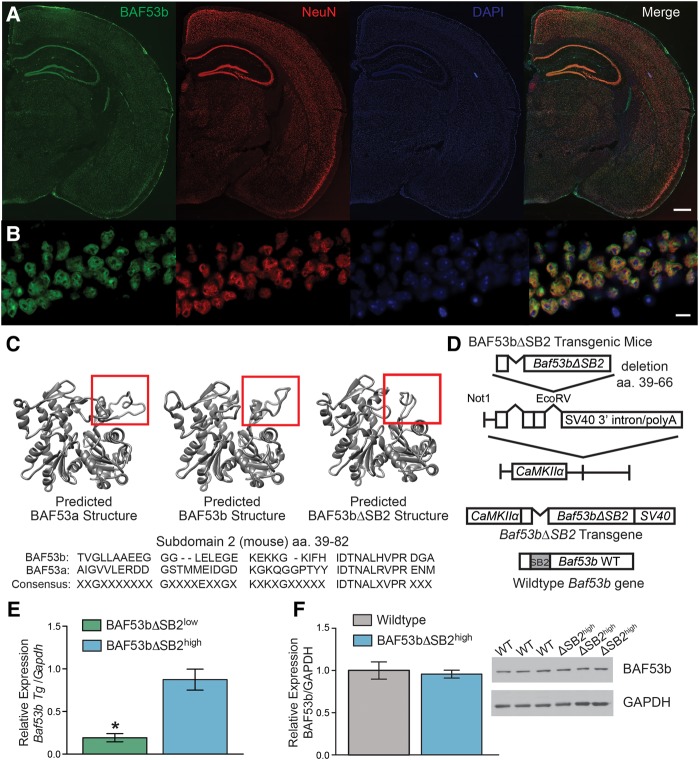
Figure 1.
Characterization of BAF53bΔSB2 transgenic mice. (A) The 4× scans of BAF53b expression in adult (12 wk) mouse brain. BAF53b expression is shown in green, NeuN expression in red, and DAPI in blue. The merged image contains all three channels. Scale bar 500 microns (B) Same panels as A but at 63× in dorsal hippocampal CA1. Scale bar 10 microns. (C) Predicted BAF53a, BAF53b, and the BAF53bΔSB2 mutant protein structure models simulated by comparison to know crystal structures for Actin using Modeller (Eswar et al. 2006). Below is shown the amino acid sequence for subdomain 2 (SB2) of BAF53b aligned to BAF53a. Note the lack of consensus across the domain. (D) BAF53bΔSB2 transgene construction. Amino acids 39–66 of the SB2 domain were deleted from wild-type BAF53b cDNA to make BAF53bΔSB2. This sequence was cloned upstream of the SV40 intron and polyadenylation signal and downstream from the 8.5 kb mouse CaMKIIα promoter. This construct was used to generate BAF53bΔSB2 transgenic mice. (E) Quantitative RT-PCR was performed with transgene specific primers to measure transgene expression in dorsal hippocampus of two independently derived lines of BAF53bΔSB2 mice (n = 9–10 animals per group; Mann–Whitney U = 4.00, P < 0.001). (F) Endogenous BAF53b expression in the dorsal hippocampus of BAF53bΔSB2high (n = 12) and wild-type (n = 12) littermates does not significantly differ (Mann–Whitney U = 56.00, P = 0.371). Mean (±SEM).

