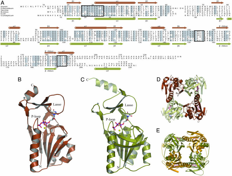
Fig. 1.
Structures of hTK1 and Uu-TK. (A) Structural alignment of the sequences of TKs from human (P04183), Dictyostelium discoideum (AAB03673.1), Vaccinia virus (AAB96503.1), B. cereus (ZP_00241105.1), E. coli (NP_287483.1), and U. urealyticum (U. parvum) (NP_078433). Secondary structure elements for hTK1 are shown above the alignment in brown, and those for Uu-TK are shown below in green. The P loop and the two zinc coordinating sequences are boxed. (B) Subunit structure of hTK1 with dTTP colored according to atom type. Mg2+ is shown in yellow, and Zn2+ is shown in gray. (C) Subunit structure of Uu-TK with dTTP, Mg2+, and Zn2+ shown in same colors as in B. hTK1 and Uu-TK are tetramers. (D and E) hTK1 (D) and Uu-TK (E) tetramers shown in different views. The C-terminal helix in Uu-TK interacts with a helix on the adjacent monomer.

