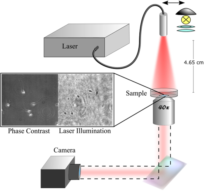
Figure 6. Setup of laser irradiation system.

Phase contrast images were taken under the microscope’s halogen lamp. During irradiation, the sample was imaged under laser illumination (without the microscope’s halogen lamp). The light source was a fiber coupled diode laser coupled to a multimode fiber with a built-in beam homogenizer.
