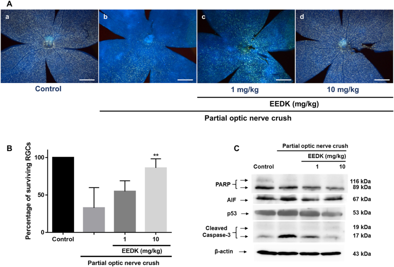
Figure 6. Effect of EEDK on RGC survival and expression of apoptosis-associated proteins in PONC-induced mice.
Forty mice (6-week-old males) were randomly divided into 4 groups (n = 8 mice per group). (A) Representative fluorescence images of retrograde-labelled RGCs in PONC-induced mice: (a) control mice, (b) vehicle (PONC only), (c) 1 mg/kg EEDK-treated group (with PONC), and (d) 10 mg/kg EEDK-treated group (with PONC). Scale bar = 500 μm. (B) The bar graph shows quantitative analysis of the RGC survival rate (%) (**p < 0.01, mean ± S.E.M.) (C) Expression of apoptotic protein levels (PARP, AIF, p53, and cleaved caspase-3) in mouse retinas after damage, with or without PONC.

