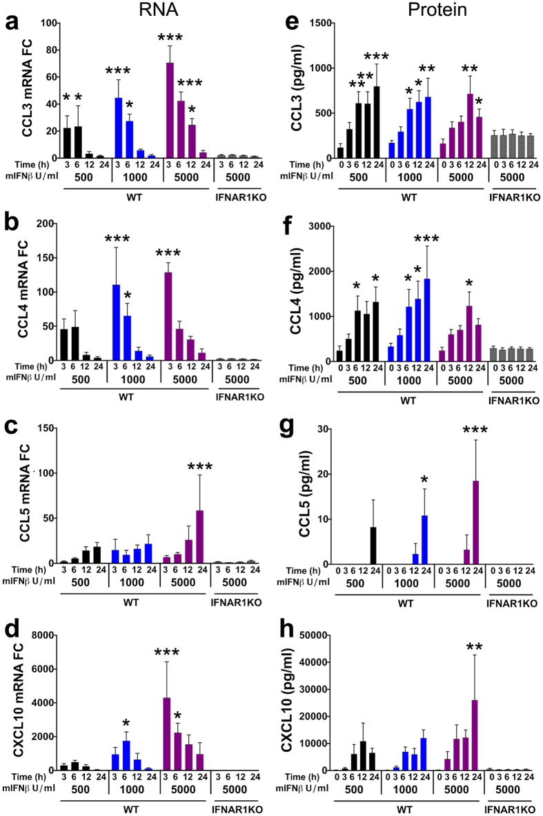
Figure 3. IFNβ stimulates expression of interferon-stimulated genes (ISG) in mixed neuronal-glial cerebrocortical cell cultures.
Mixed neuronal-glial cerebrocortical cultures from WT or IFNAR1KO mice were incubated with IFNβ in increasing doses (from 500 to 5,000 U/ml) or BSA/PBS vehicle control for 0, 3, 6, 12 and 24 h. Total RNA was extracted from cell lysates, analyzed by qRT-PCR and normalized to GAPDH expression levels. (a–d) RNA expression is shown as fold change (FC) in relation to vehicle treated controls which were defined as baseline activity. (e–h) Time course for protein expression measured in cell-free supernatants for CCL3, CCL4, CCL5 and CXCL10 using a commercially available multiplex assay as described in Methods. Baseline protein expression in vehicle treated cell cultures is represented as 0 h time point. Values are mean ± s.e.m.; n = 3–5 independent experiments per ISG; ***p < 0.001, **p < 0.01, *p < 0.05 by ANOVA with Fisher’s PLSD post hoc test. For clarity, the significance is only indicated for differences between treatments and baseline within each experimental group.

