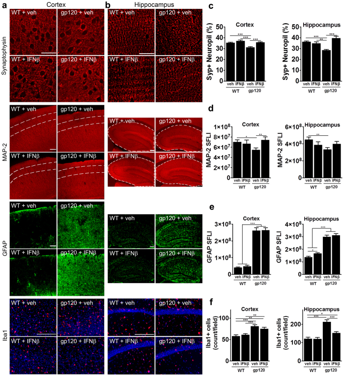
Figure 5. Intranasally administered IFNβ prevents neuronal damage in HIVgp120tg mice.
Three to four month-old HIVgp120tg and WT littermate male mice received intranasal recombinant murine IFNβ (50,000 U/25 g bodyweight) or vehicle once a week for four weeks and afterwards, the brains were assessed for neuronal damage and glial activation. Representative images of frontal cerebral cortex (a) and hippocampus (b) immunolabeled for neuronal synaptophysin (SYP; cortex layer III, hippocampus CA1) deconvolution microscopy; scale bar, 40 μm) and MAP-2 (cortex layer III (area between dashed lines) and hippocampus) fluorescence microscopy; scale bar, 100 μm), astrocytic GFAP (scale bar, 100 μm) and microglial Iba1 (red, DNA in blue, scale bar, 100 μm). (c–f) Quantification of microscopy data obtained in frontal cortex and hippocampus of sagittal brain sections of four to five month-old HIVgp120tg mice and controls treated with IFNβ or vehicle: (c) SYP in percent positive neuropil, (d) MAP-2 immunoreactivity as sum of fluorescence intensity (SFLI; arbitrary units); (e) fluorescence signal for astrocytic GFAP; (f) quantification of Iba1+ microglia (counts/microscopic field). Values are mean ± s.e.m.; ***p < 0.001, **p < 0.01, *p < 0.05; ANOVA and Fisher’s PLSD post hoc test; n = 4–5 animals per group/genotype.

