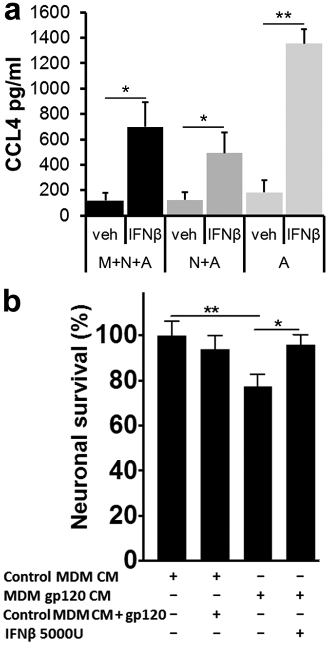
Figure 8. Interaction of IFNβ with neurons and astrocytes suffices to protect against neurotoxicity of HIVgp120-stimulated macrophages.

(a) Cerebrocortical cultures from mice were prepared to either contain microglia, neurons and astrocytes (M + N + A) or were depleted of microglia (N + A) or neurons and microglia (A). Complete and depleted cell cultures were incubated with mIFNβ (5,000 U/ml) or BSA/PBS vehicle control for 0, 3, 6, 12 and 24 h and concentrations of CCL4 were measured in cell-free supernatants using a commercially available multiplex assay as described in Methods. Maximum concentrations were reached in samples of 12 to 24 h mIFNβ exposure and compared to vehicle-treated, baseline samples. Values are mean ± s.e.m.; n = 3 independent experiments; *p < 0.05, student’s t-test. (b) Microglia-depleted rat cerebrocortical cultures were exposed for 24 h to 50% cell-free conditioned media (CM) from human MDM in the presence or absence of human IFNβ (5,000 U/ml). MDM were previously stimulated for 24 h with HIV-1 gp120BaL (MDM gp120 CM) or vehicle (MDM CM). Following the incubation the cells were fixed and permeabilized. Neurons were immunolabeled for neuronal MAP-2 and NeuN and nuclear DNA was stained with H33342. Neuronal survival was assessed using fluorescence microscopy and cell counting as described in Methods. Values are mean ± s.e.m.; n = 2 independent experiments, with 4–8 replicates and an average of 4,000 cells counted per condition; **p < 0.01, *p < 0.05 by ANOVA with Fisher’s PLSD post hoc test.
