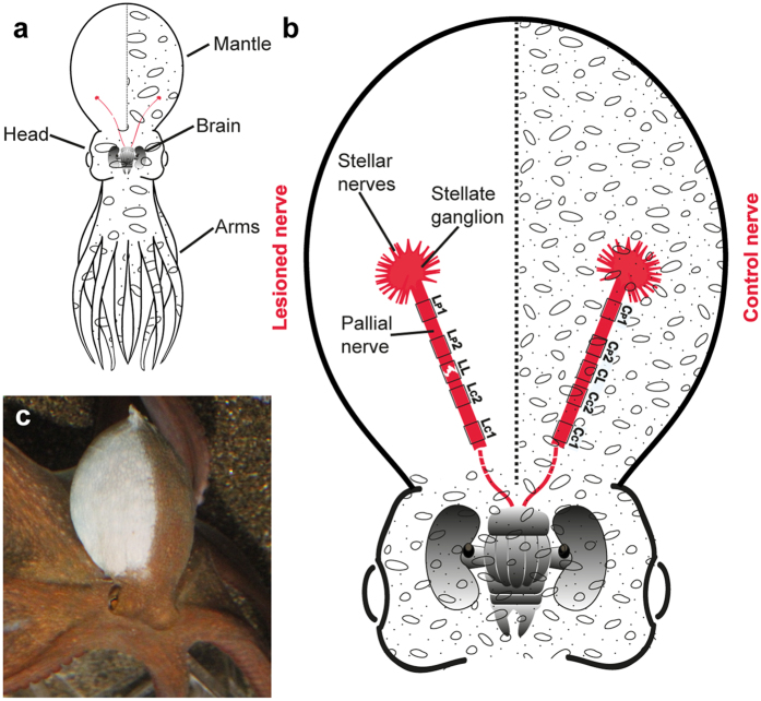
Figure 1. Main features of the anatomy of Octopus vulgaris in relation to the pallial nerve, and effects of its lesion.
(a) General anatomy of O. vulgaris. In red the two pallial nerves are visible rising from the posterior part of the brain, each of them directed toward the ipsilateral side of the mantle. (b) Enlargement of head and mantle to show the two pallial nerves ending each into a stellate ganglion. For each nerve five areas are identified for the purpose of this study, and used as target for imaging and subsequent analysis: a distal and a proximal area in the central stump (respectively identified as LC1 and LC2), similarly in the peripheral stump (LP1 and LP2) of the transected nerve. Analogous areas were identified in the control nerve, i.e.: the one corresponding to the lesion site (CL), a distal and a proximal area in the central ‘stump’ (CC1, CC2), and at the level of the peripheral ‘stump’ (CP1, and CP2). (c) Right pallial nerve transection causes complete paling of the mantle at the level of the denervated side due to loss of neural control of the chromatic and textural components of body patterns. Body parts are easily recognizable (see a,b).

